T.R. Jenkyn
Contact
Department of Mechanical & Materials Engineering
Thompson Engineering Building,
Room TEB 367
Western University
Tel: 519-661-2111 ext. 88339
tjenkyn@uwo.ca
Projects
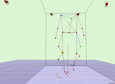
I am Co-Director of the Wolf Orthopaedic Biomechanics Laboratory (WOBL) and Director of the Wolf Orthopaedic Quantitative Imaging Laboratory (WOQIL), both of which study orthopaedic patients at the Fowler Kennedy Sport Medicine Clinic (FKSMC). The WOQIL is unique in Canada in that it combines 3D bi-planar fluoroscopic imaging with optical motion analysis, a force instrumented treadmill and pressure measurement. My overall goal is to quantify in-vivo motion and loading of bones within articular joints to identify the biomechanical causes of osteoarthritis and other musculoskeletal disorders. I am active in four areas: 1) 3D fluoroscopic radiostereometric analysis (f-RSA) for use in biomechanics, 2) motion analysis of the joints of the foot, 3) optical gait analysis of knee osteoarthritis, and 4) biomechanical analysis of elite and recreational sport.
3D Fluoroscopic Radiostereometric Analysis (f-RSA)
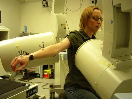
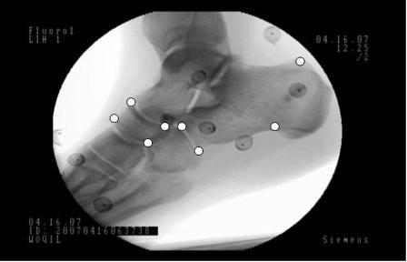
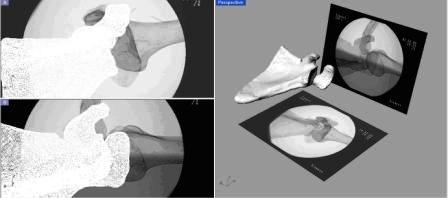 Funded by CFI ("Abnormal arthrokinematics and osteoarthritis"), the WOQIL was set up. This novel facility uses bi-planar fluoroscopic radiostereometric analysis (f-RSA) to resolve moving 3D bone position and orientation in-vivo to accuracies of +/-40 microns and 0.1 degrees. There is no error arising from skin motion, as is the case with optical motion analysis. Specifically I am using this system to study scapular-humeral kinematics in the shoulder, foot bone motion within footwear and joint kinematics in the osteoarthritic knee. This system can be used with or without implanted marker beads, allowing for healthy subjects to be studied.
Multi-segment Foot Model and Foot Joint Motion
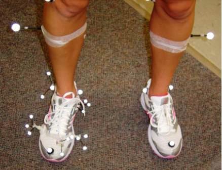 I developed a multi-segment kinematic foot model for use with optical motion capture. The model tracks individual foot segments during dynamic activities. The model can be used with or without shoes, orthotics or bracing, allowing the influence of these interventions to be quantified. This model has been used by the Nike Sport Research Lab (Nike Inc, Beaverton, OR, USA). This model, along with f-RSA, currently forms the basis of my research into running shoe design, orthotic development and intrinsic muscle strengthening.
Injury causation biomechanics
 I use f-RSA and optical motion capture to quantify the loads (forces and torques) acting on the bones and soft tissues of human subjects during various simulated injurious activities. These include slip-trip-fall motions, dislocation and tears of the shoulder, head and neck inertial injuires and injuries to the face. This research clarifies the biomechanical mechanisms of acute and chronic injuries to aid in the differential diagnosis of injury causes after the incident. As a biomechanical engineer, I am also active in the design and development of novel protective equipment and garments for the avoidance of musculoskeletal and brain injuries.
Gait Analysis of Knee Osteoarthritis
 With my Co-Directors, I have contributed to the clinical understanding of the high tibial osteotomy (HTO) as a treatment to slow the progress of knee osteoarthritis. Funded by a CIHR University-Industry partnership (with Arthrex, Inc, FL, USA) and recently renewed for another 5 years ("High tibial osteotomy: Lessening the load"), we have shown an excellent 2-year outcome and are examining the 5-year outcome. Our database holds more than 600 patients and 1200 gait analyses.
Elite and Recreational Sport Biomechanics
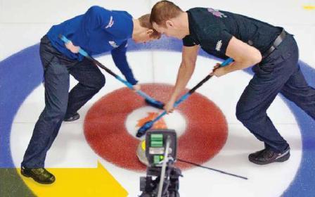 I have active research in two Olympic sports: rowing and curling. The Canadian Olympic Committee, Own the Podium 2010 Program funded my investigation into sweeping in curling. This resulted in a patent for a new broom that was used by our teams at the Vancouver 2010 Games. This included training camps using optical motion capture and infrared imaging in-situ. By agreement, I am unable to publish this work until the summer of 2010. My research on Olympic rowers has been lab-based and water-based and has included consultation with national level coaching staff.