Current Projects
Contact Us
Email: rwilling@uwo.ca
Lab Locations
Thompson Engineering Building (TEB 306)
University Hospital (CLL-100)
Find out more about the capabilities of these labs on the Lab Facilities / Equipment page.
Biomechanical Analysis of Knee Ligaments (intact, reconstructed and replacement)
Introduction: Although the posterior cruciate ligament (PCL) is the strongest ligament in the knee, PCL injuries still account for between 3% and 20% of knee injuries. PCL injuries can happen during sports when athletes land on the ground with their knee flexed or during car accidents. PCL injuries usually happen in conjunction with injuries to other stabilizing structures of knee. Whilst it is commonly accepted that the posterolateral structures need to be addressed surgically in high grade PCL injuries, it is unclear which medial structures need to be addressed and when. The posterior oblique ligament (POL) and deep medial collateral ligament (dMCL) are two medial ligamentous structures whose behavior in the PCL-deficient knee are not well characterized. The hypotheses of this study are: 1) by dissecting either of these two structures in combination with the PCL, more posterior tibial translation will be observed than in an isolated PCL injury, 2) by characterising these structures with multiple bundles instead of one bundle we can more accurately simulate knee kinematics.
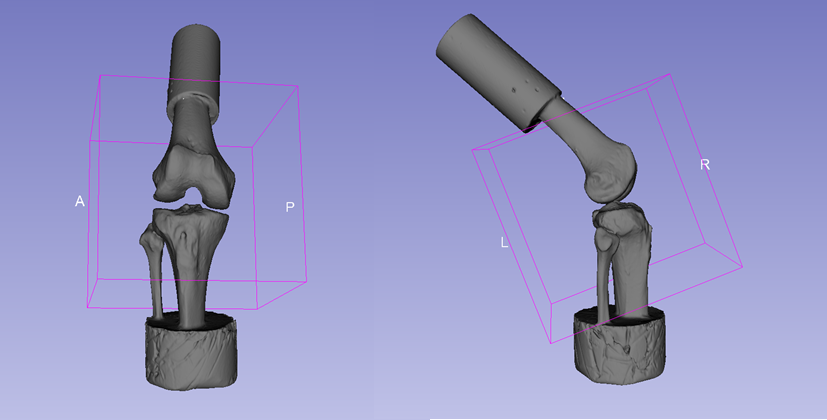
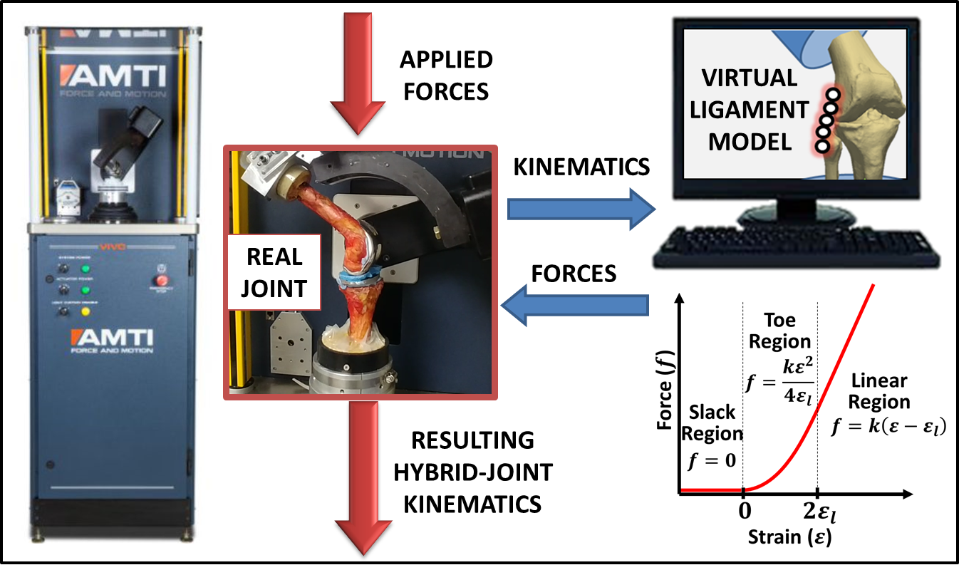
Methods: intact cadaver knees are being used in this study. Knees are potted, aligned and mounted onto a VIVO joint motion simulator (Advanced Mechanical Technologies, Inc.) using custom-designed fixtures which allows repeatable positioning of specimens. Once installed, specimens are subjected to a baseline loading scenario which consisted of prescribed flexion from 0 to 90 degrees with a 100 N compression force applied along the long axis of the tibia; all remaining degrees of freedom are unconstrained. The resulting joint kinematics represent the natural path of motion of the joint. The motion is then repeated with additional loads applied to the tibia to measure the stability of the joint, including (1) a posterior-directed force of 67 N or (2) 2.5 Nm internal and external torques. The tests are repeated following sequential ligament dissections. The PCL is first dissected arthroscopically, followed by dissection of either the POL or dMCL performed in a randomized order (3 POL first, 3 dMCL first). The dMCL and POL are accessed by temporarily exposing the medial side of the knee through an incision which is stapled closed after ligament dissections. All data a processed in order to calculate the kinematics of the tibia relative to the femur. The knee will go through resulting kinematics from intact and PCL-deficient stages and the joint reaction forces are being recorded. Using superposition principle, we are measuring the force contribution of each ligament in both intact knee and PCL-deficient knee.
Current Progress: Currently, we have collected translations and joint reaction forces for 13 knees. Next step in our experiment would be tuning virtual ligaments for individual knees and replacing dissected ligaments with these virtual ligaments, and comparing the kinematics with kinematics of knees when they are intact. Hitting the limits of range of motion of the VIVO has been one of the most occurring challenges in this experiment. Other challenge has been damaging the structures during experiments.